

About
Scopio Labs is an enterprise-grade AI platform purpose-built for clinical hematology. It digitizes the microscopy workflow end-to-end, offering full-field imaging at 100x resolution for both peripheral blood smears and bone marrow aspirate slides. Rather than manually scanning 100 cells at a microscope, lab teams can now leverage autonomous AI to analyze thousands of cells per slide, dramatically improving detection rates for rare cell types such as blasts, dysplastic cells, and plasma cell dysplasias. The platform's AI-powered Complete Blood Morphology (CBM) solution automates differential cell counting and generates structured reports, reducing inter- and intra-user variability that has long plagued manual hematology reviews. Full-field digital scans allow clinicians and hematopathologists to navigate freely across the entire slide, mimicking and surpassing the experience of bench microscopy. Scopio's remote-access architecture lets distributed teams—including multi-hospital networks—share expertise, review slides, and collaborate in real time over a secure network without any physical slide transport. This addresses critical staffing shortages and rising testing volumes in modern clinical labs. Scopio Labs is designed for large reference laboratories, academic medical centers, and multi-site hospital systems seeking to standardize morphology quality, reduce reliance on on-site staff, and future-proof their hematology workflows with scalable digital and AI capabilities.
Key Features
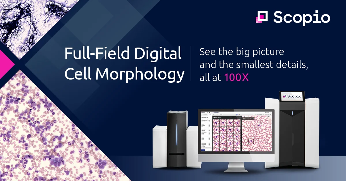
- Full-Field Digital Imaging at 100x: Captures complete, high-resolution digital scans of blood and bone marrow slides at 100x magnification, enabling free navigation across the entire slide just like a physical microscope—but remotely.
- AI-Powered Cell Differential (CBM): Complete Blood Morphology autonomously analyzes thousands of cells per slide, generating structured differentials and final reports while reducing human variability in cell classification.
- Remote Access and Collaboration: Enables pathologists, hematologists, and lab staff to review, annotate, and consult on slides from any location over a secure network, eliminating the need for physical presence at the microscope.
- Bone Marrow Aspirate and Peripheral Blood Smear Support: Dedicated applications for both peripheral blood smear review and bone marrow aspirate analysis, covering the full scope of clinical hematology morphology workflows.
- Standardization Across Multi-Site Networks: Enables hospital systems and reference labs with multiple sites to unify morphology quality and share expertise seamlessly, reducing site-to-site variability in diagnostic outcomes.
Use Cases
- Clinical hematology labs replacing manual light microscopy with fully digital, remote slide review workflows
- Multi-hospital systems standardizing morphology quality and sharing hematopathology expertise across all sites from a centralized location
- Academic medical centers conducting high-volume bone marrow aspirate and peripheral blood smear analysis with AI-assisted differentials
- Reference laboratories managing rising test volumes and critical staffing shortages by enabling remote and asynchronous pathologist review
- Hematopathologists consulting on complex cases involving blasts, multiple myeloma, or plasma cell dysplasias by freely navigating full-field digital slides
Pros
- Dramatically More Cells Analyzed: Analyzes thousands of cells versus the traditional 100-cell manual differential, significantly improving statistical confidence and detection of rare cell types.
- Fully Remote Workflow: Eliminates slide transport and on-site requirements, allowing expert consultation and sign-out from anywhere—critical for labs facing staffing shortages.
- Proven Diagnostic Image Quality: Widely endorsed by leading hematopathologists and academic medical centers as providing image quality equivalent to or better than direct light microscopy.
- Faster Turnaround Times: Delivers up to 60% faster peripheral blood smear review turnarounds through AI-assisted differential counting and streamlined digital workflows.
Cons
- Enterprise Pricing: Scopio Labs is priced for hospital systems and large reference labs, making it inaccessible for smaller independent labs or individual practitioners.
- Hardware Dependency: Requires Scopio's proprietary scanning devices to capture full-field images, meaning existing lab equipment cannot be repurposed for the platform.
- Regulatory and Integration Complexity: As a clinical diagnostic tool, deployment involves regulatory compliance, LIS integration, and institutional validation workflows that can extend implementation timelines.
Frequently Asked Questions
What types of slides can Scopio Labs analyze?
Scopio Labs supports both peripheral blood smear slides and bone marrow aspirate slides through dedicated applications built for each specimen type.
Does Scopio Labs replace the need for on-site pathologists?
Scopio enables remote review and consultation, so hematopathologists and clinicians no longer need to be physically present in the lab. However, trained professionals still review AI-generated differentials before final sign-out.
How does the AI differential compare to a manual differential?
Scopio's AI analyzes thousands of cells compared to the standard 100-cell manual differential, providing greater statistical confidence and improved sensitivity for rare or abnormal cell types.
Is Scopio Labs suitable for multi-hospital networks?
Yes. Scopio is specifically designed for multi-site hospital systems and large reference labs, allowing centralized expertise and standardized morphology quality across all connected facilities.
What imaging resolution does Scopio provide?
Scopio captures full-field digital images at 100x magnification, the standard oil-immersion resolution used for blood and bone marrow morphology, delivering images that match or exceed what is visible through a traditional light microscope.